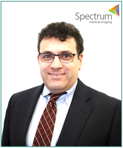
Interview with Dr. Daniel Moses, Partner @ Spectrum Medical Imaging
Welcome to the third post of our interview series. Given AI and machine learning is pervading many industries and professions, we thought we’d reach out to a few experts in various industries to find out more about their experience to date. AI has started to have a real impact in medical imaging, so for this instalment, we managed to catch up with Daniel Moses, partner at Spectrum Medical Imaging, radiologist and PhD in Computer Science.

VL: Tell us a little bit about yourself.
DM: I am a subspecialist radiologist in body imaging (which includes the chest, abdomen, pelvis and heart). I have completed two clinical fellowships in Thoracic and Body Imaging at New York University. I am working in the public and private sector. I also have a background in Science (majors in mathematics, physics, biochemistry and physiology, honours in pure mathematics) and Engineering (Master of Engineering Science in Biomedical Engineering and PhD in Computer Science and Engineering). I am involved in clinical practice and research. My research interests include MR and CT imaging of the pancreas, prostate and heart. I also have a keen interest in medical imaging analysis and artificial intelligence applied to medical imaging problems. I am passionate about bridging the gap between engineers and clinicians and have recently take up a role as Medical Director - Research Imaging NSW at UNSW which allows me to work with both groups.
VL: How do you think radiology practice is going to change in the coming years?
DM: I think the first change will result in certain tasks becoming more automated and efficient. These tasks included processes involving workflow and the reporting of cases. For example intelligent systems will analyse the unreported imaging cases available and send reporting studies to those radiologists most capable and efficient in analysing cases in that area (e.g. oncology of the chest). IT systems will also make generating a radiology report easier by automatically generating report templates which are prepopulated with measurements of organs and lesions - which is currently done manually. This will increase speed and decrease transcription errors. Computer systems will also make it much easier to correlate with other imaging studies: either prior studies or concurrent studies in a different modality (such as ultrasound). This will make characterisation of pathology/diseases a lot easier and more accurate.
A little while later the ability for algorithms to screen the medical images themselves (via CNNs etc) for abnormalities will improve to the level that they will be used routinely in clinical practice. I think these will be used as assistants for the reporting radiologists, allowing them to read studies more efficiently. They won’t take over the reporting.
Interventional radiologists will also be assisted by robotic systems in performing biopsies and patient treatments like arterial embolisation and stent placement.
The ability to extract quantitative imaging bio-markers (QIBs) from image data is very important. Measuring a feature of an image reliably and reproducibly will let us discover features that can predict how aggressive a disease will be and how well it is responding to therapy. This is especially important in cancer. The new field of radiomics attempts to generate thousands of features from medical images and then find the ones that are important. When radiomics combines with genomics and other clinical data it is possible to “personalise” a patient treatment to that particular cancer, thereby increasing their chance of survival. This is known as personalised medicine. This is currently a field under intense research and I believe will become increasingly important. AI systems are ideal for helping navigate radiomics and finding features.
It is going to take quite some time for artificial intelligence systems to perform some of the more complex tasks that radiologists perform, including synthesizing complex findings into a unifying diagnosis, planning the best path to investigate a patient with confounding findings, participating in multidisciplinary teams, optimising imaging protocol and running a complex clinical service.
VL: Absolutely. Augmentation rather than automation, at least in the foreseeable future. In terms of what’s happening now, what do you believe are some of the more exciting AI applications you’ve seen?
DM: I think there is great software available for task optimisation including worklist management and reporting. Some of the new AI systems can essentially remove typing altogether.
AI applications that perform menial tasks, such as identifying lung nodules, and calculating the ejection of the heart are becoming very accurate, outperforming human in speed and reproducibility. These are very exciting.
The new AI breast systems which look at mammograms are also very good and many are being rolled out in large services.
Also, deep learning methods are now replacing many of the computationally intensive algorithms used to generate medical images from raw data. They can often speed up the process many-fold resulting in much more efficient scan times. These are already being rolled out into clinical practice and are having an impact. An example is the reconstruction of CT images from low dose raw imaging data.
VL: Amazing stuff. So are you or Spectrum planning on investing / working with / exploring AI and machine learning? What have you done so far?
DM: Yes - we are participating in radiomics research looking for imaging features in cancer that have value in predicting tumour behaviour and treatment response. We are also looking at other applications such as screening for vertebral fractures and assessing for abnormalities of the inner ear.
VL: Have you worked with any data scientists or other AI practitioners? What has been your experience?
DM: Yes. I have worked with computer scientists in research projects. Although very skilled in the algorithmic development, I have found that they don’t have knowledge of the medical and radiological processes we are modelling and therefore lack insights into what is important in the data. That is why a team approach is paramount. Even better, we need to educate people more broadly so that they understand both the computing and radiology well.
VL: Couldn’t agree more. In general then, how do you think other radiologists and medical imaging companies could be better prepared for technological change?
DM: I think understanding the technologies and the science behind them is paramount for radiologists. Knowing, for example, about biases in machine learning is very important. Medical imaging companies need to establish ties with clinicians who understand the important problems that need to be solved.
VL: Are there any particular companies you’re watching in this space?
DM: Yes - the large vendors that sell medical imaging equipment (e.g. GE, Siemens). They have huge resources and are already investing in massive deep learning infrastructure. They also have access to masses of imaging data and have entry to front line clinical practice via their products which include imaging machines and data analysis packages.
VL: Definitely worth keeping an eye on. You mentioned biases in machine learning - more broadly, regarding explainability - how much do you care about how a model works, or is it only important that it works sufficiently?
DM: I think the ability to explain how the algorithm is working is important although not always completely possible. By understanding what it is doing (in some sense or another) you can glean insights that may help you improve the process and unveil potential biases in the system.
A classical example of biases is when researchers wanted to screen resumes for the best programmers. They trained their algorithms using data on all of the existing programmer population in Silicon Valley at successful companies. The problem is that the algorithm’s best selection criteria was to exclude any CV that had grammar that suggested it belonged to a female. This led to no females being considered due to the fact that there were very few in the training data. Using this AI system would result in decreased diversity which in my opinion would be to the detriment of the employers. It has been constantly shown that diverse teams do better than non-diverse teams when solving problems by the sheer increase in diversity of perspectives and approaches that they consider.
VL: That’s a great example. It’s also exactly what our software Oversight.ai seeks to mitigate - but it’s far from simple. Finally, any parting words?
DM: I think the “specific AI” applications will progress for a while and then plateau. We are nowhere near creating a general AI. I think this will eventually happen and that will be the real game changer.
VL: It’s certainly an interesting debate. Well, thank you so much for sharing these insights with us, I know I’ve learnt a lot, and I’m sure our readers will too!
To find out more about Daniel and Spectrum, check out spectrumradiology.com.au.
